
iCare COMPASS
iCare COMPASSiCare COMPASS Automated Perimeter with active Retinal TrackingKey features*Standard automated perimetry*Active retinal tracking compensating for poor patient fixation in real-time*Auto-fo
- Weight::
- Brand ::
- No.::
iCare COMPASSiCare COMPASS Automated Perimeter with active Retinal TrackingKey features*Standard automated perimetry*Active retinal tracking compensating for poor patient fixation in real-time*Auto-fo
iCare COMPASS Automated Perimeter with active Retinal Tracking
*Standard automated perimetry
*Active retinal tracking compensating for poor patient fixation in real-time
*Auto-focus — no trial lens needed
*Hygienic design
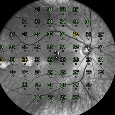
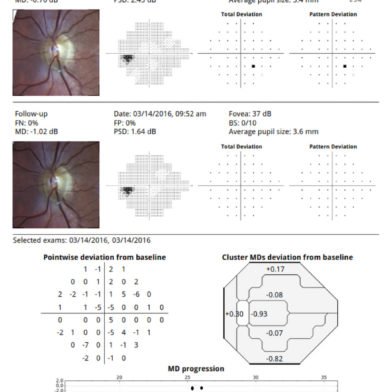
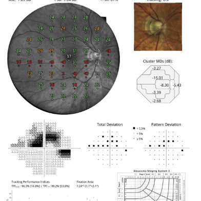
*Illustrative fixation analysis; fixation area and plot
*High-resolution confocal TrueColor imaging of the retina
*No dilation of pupil needed, the patient can blink freely and the test can be suspended at any time without data loss
*Ease of use & minimal operator training
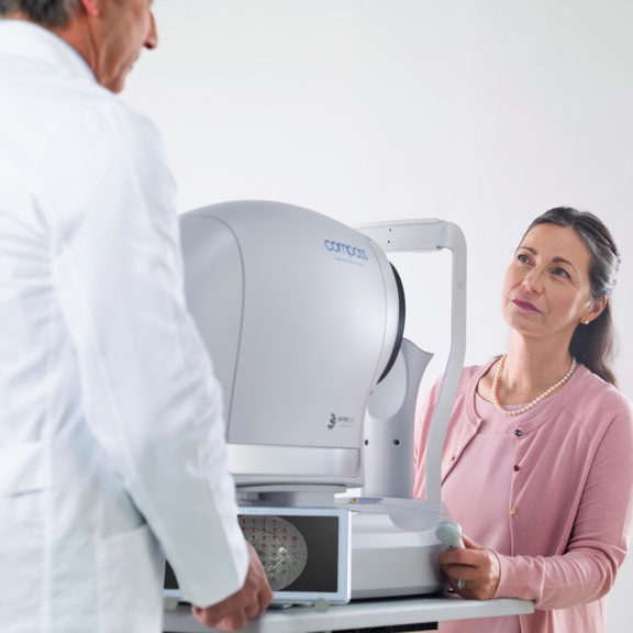
iCare COMPASS combines visual field tests, fixation loss correction by a real-time retinal tracker and confocal TrueColor fundus imaging taking visual field analysis to the next level. With touch screen, auto-alignment, non-mydriatic, easy-to-disinfect, and trial lens-free operation, iCare COMPASS is patient-friendly and easy to use, saving time and helping in improving clinical performance.
iCare COMPASS is the first automated perimeter that can perform standard visual field tests using a real-time retinal tracker while delivering ultra-high resolution confocal TrueColor fundus images at the same time.
Now you have a chance to get improved results easier by replacing your existing perimeter with iCare COMPASS. We are offering a trade-in campaign until the end of December 2021.
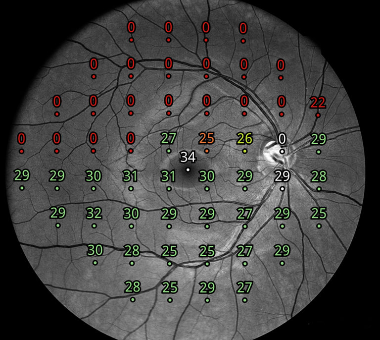
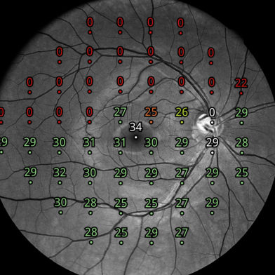
Fundus-controlled perimetry is a technique that images the retina during visual field testing, enabling a reliable correlation to be made between visual function and retinal structure. Retinal tracking is at the heart of this function.
Continuous, automated, tracking of eye movements by infrared scanning of the retina provides active compensation for fixation losses, with perimetric stimuli automatically repositioned prior to and during projection based on the current eye position. This ensures an accurate match between function (i.e. retinal sensitivity values) and structure (fundus image) thanks to the compensation for eye movements resulting in the reduction of motion artifacts.
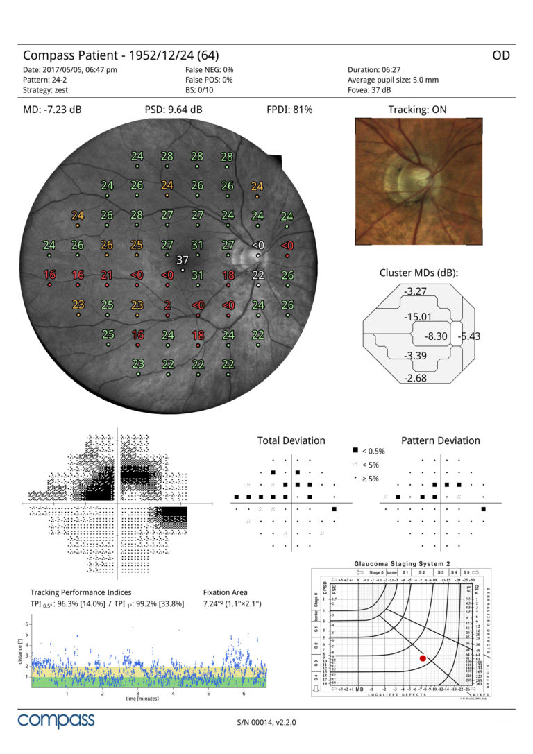
Automated visual field test with TrueColor Confocal imaging
As a perimeter, iCare COMPASS offers full compatibility with standard 24-2, 30-2 and 10-2 visual field testing containing age-matched databases of retinal sensitivity in normal subjects. For the first time in a visual field test, iCare COMPASS provides 60° confocal images of the retina in TrueColor, Infrared and Red-free. iCare COMPASS is also the first perimeter to provide high resolution stereo viewing for a better assessment of the optic nerve head.
iCare COMPASS is the first fundus-controlled perimeter that can perform standard visual field tests, while delivering ultra-high resolution confocal TrueColor fundus images simultaneously.
With iCare COMPASS Automatic Refraction Correction, the Auto-focus, there is no need for trial lenses. This not only reduces examination time but avoids lens rim artefacts and improves patient flow and comfort. The test can be suspended at any time without data loss. The touch screen interface via high resolution integrated tablet and the all-in-one design (no external PC) with 1 Ethernet and 3 USB Ports make the device easy to operate. iCare COMPASS also offers embedded capabilities for network connectivity that allows remote viewing.
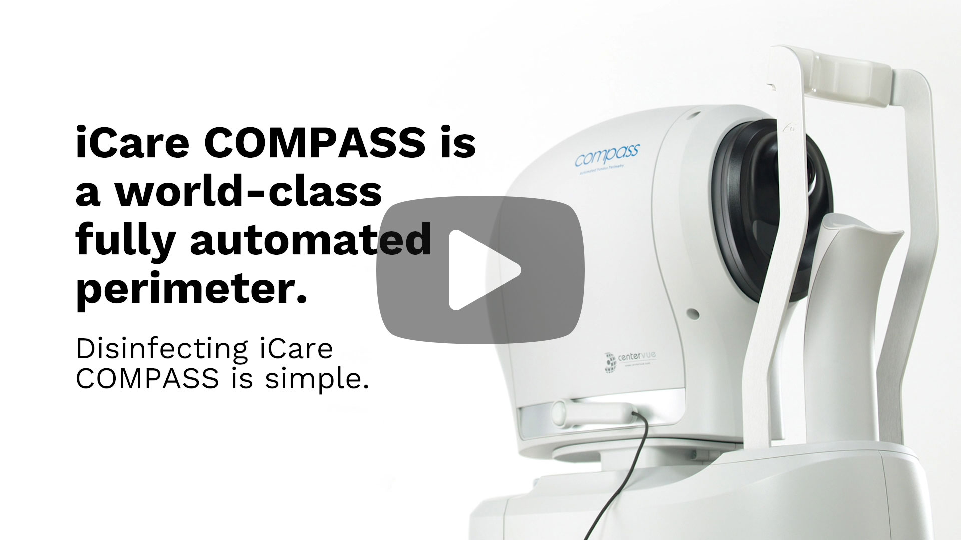
The design of iCare COMPASS offers an additional benefit of being easy to clean between patients due to the smooth, narrow and convex front of the device: no bowl and trial lens assembly. All iCare COMPASS surfaces can be disinfected with an alcohol wipe. Watch the video for more information.
Fundus Perimetry
Projection field30° (radius)
Background luminance31.4 asb
Maximum luminance10000 asb
Dynamic range0 - 50 dB
Stimulus sizeGoldmann III (26”)
Stimulus duration200 ms
Test StrategiesZEST, 4-2, Supra-threshold
Threshold tests30-2, 24-2, 10-2
Fixation control25 Hz automated retinal Tracking
Foveal threshold testing
Automatic pupil size measurement
Fundus Imaging
Field of view60° (diameter) [Center of Eye Angle of 90° (H)]
Bi-focal Stereo Image of the ONH
Sensor resolution5 Mpixel (2592x1944)
Light sourceinfrared (825-870 nm) and white LED (440-650 nm)
Imaging modalitiescolor, infrared, red-free
Resolution17 microns
Dimensions
Weight25 Kg
SizeH 620 X W 590 X D 360 mm
Electrical requirements
Power100-240 VAC, 50-60 Hz
Consumption80 W
Other Features
Automatic operationauto-alignment, autofocus, auto-retinal tracking, auto-pupil tracking, auto-exposure, auto-capture
Non-mydriatic operationminimum pupil size 3 mm
Working distance28 mm
Auto-focusing adjustment range-12D to +15D
Fixation targetprogrammable, internal
User interfaceTablet operated, with multi-touch, color display
Ethernet connection through device
DICOM supportmodality worklist
Hard diskmodality worklist