iCare DRSplus
iCare DRSplus TrueColor confocal fundus imaging systemKey featuresTrueColor Confocal TechnologyMultiple imaging modalities including Red-free, external eye and stereo view imaging2.5 mm minimum pupil
- Weight::
- Brand ::
- No.::
iCare DRSplus TrueColor confocal fundus imaging systemKey featuresTrueColor Confocal TechnologyMultiple imaging modalities including Red-free, external eye and stereo view imaging2.5 mm minimum pupil
iCare DRSplus TrueColor confocal fundus imaging system
* TrueColor Confocal Technology
* Multiple imaging modalities including Red-free, external eye and stereo view imaging
* 2.5 mm minimum pupil size
* Fast, easy and fully automated operations
* Mosaic function which creates retinal panoramic views up to 80°
* Remote Viewer that allows for reviewing from devices on the same local area network
* Remote Exam feature enables executing an exam from a distance
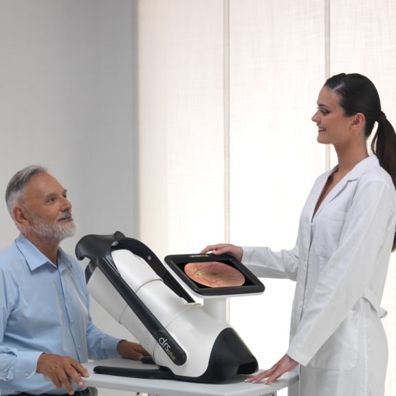
iCare DRSplus Confocal Technology speeds up exam time while ensuring a comfortable patient experience. The device permits imaging through pupils as small as 2.5 mm and this non-mydriatic capability eliminates need for dilating drops and dark environments. With reduced flash intensity for softer effect on the pupil and quick and easy patient positioning, iCare DRSplus is a patient-friendly device.

iCare DRSplus is an easy-to-use and intuitive imaging system that requires minimal staff training to obtain the highest quality images. iCare DRSplus offers live IR preview which contributes to an efficient capture workflow, while the filtering option makes image post-processing simpler. The fully-automated capabilities include auto-alignment, auto-focus, auto-exposure, auto-capture and auto-montage (optional). Quick and easy patient positioning and speedy examination saves time and makes the workflow efficient.
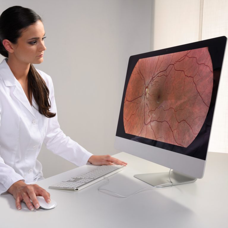
iCare DRSplus capacitive touch screen allows for easy magnification and review of images’ details. Additionally, the optional remote viewer feature makes remote data review effortless, allowing any computer or laptop on the same local area network (LAN) to review iCare DRSplus images remotely, enabling data access and detailed analysis on multiple review stations.
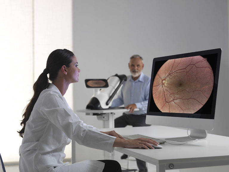
In the fight against COVID-19, healthcare professionals are required to apply special social distancing precautions to minimize the spread of the virus. We are working closely with healthcare professionals during this difficult period to provide appropriate support and tools to facilitate social distancing during patient exams.
iCare DRSplus confocal fundus imaging system uses white LED illumination to offer high-quality TrueColor images. TrueColor Confocal Technology, which is considered a standard of high image quality, provides detail-rich images with greater image sharpness, optical resolution and contrast when compared to traditional fundus camera imaging.
The fast and fully automated iCare DRSplus permits imaging through pupils as small as 2.5 mm, without need of dilation, ensuring a comfortable patient experience. This easy to use device offers the advantage of quick examination time and helps speed up workflow at clinics.
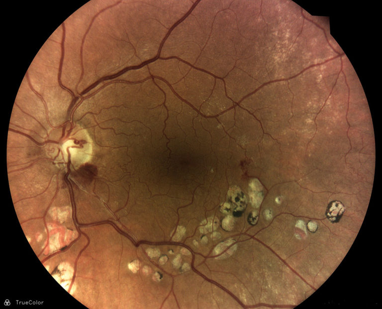
TrueColor Confocal Technology with white LED, promotes detailed 45° retinal images and allows to scan through cataract to aid clinicians in the diagnosis and documentation of ocular disease.
High resolution red-free filtering enhances visualization of retina vasculature, blue images provide improved view of the Nerve Fiber Layer (RNFL) and red channel allows light to penetrate into the deep layers of theretina. External eye imaging can document eye surface and cornea conditions. Stereo viewer technology creates improved 3D perception of the disc. The option of mosaic function automatically combines different retinal fields without user intervention, creating panoramic views up to 80°.
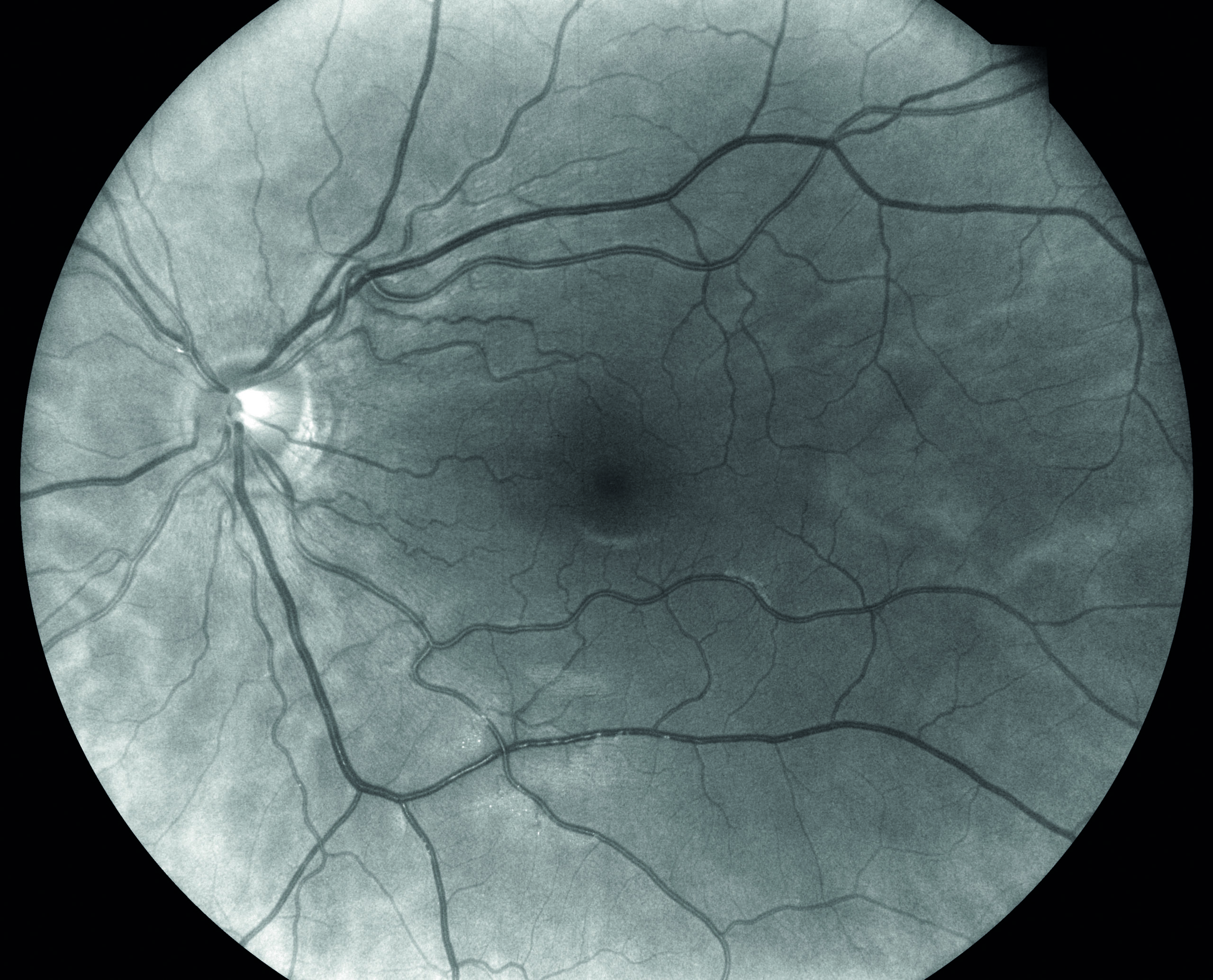
TrueColor Confocal Technology with white LED, promotes detailed 45° retinal images and allows to scan through cataract to aid clinicians in the diagnosis and documentation of ocular disease.
TrueColor Confocal Technology with white LED, promotes detailed 45° retinal images and allows to scan through cataract to aid clinicians in the diagnosis and documentation of ocular disease.
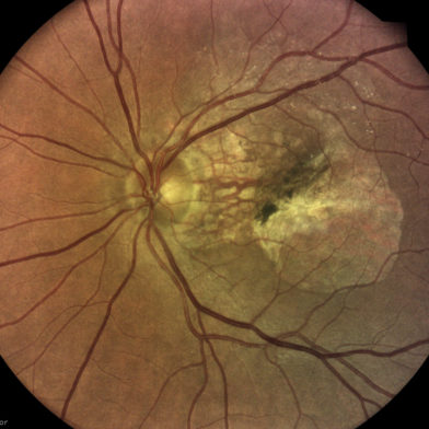
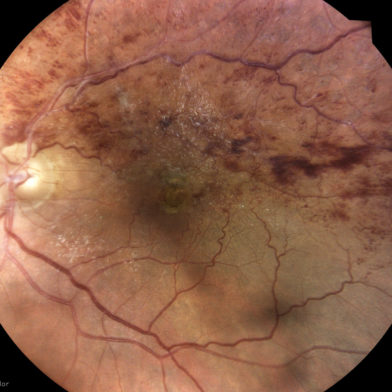
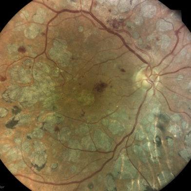
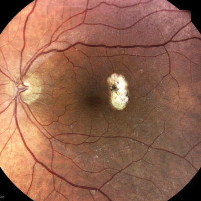
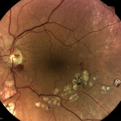
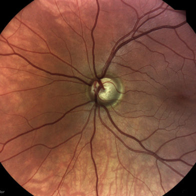
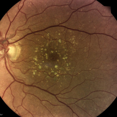

Fundus Imaging System Features
Field of ViewSingle image (W x H): 45° x 40°, Mosaic up to 9 fields (W x D): 83° x 78°
Light SourcesWhite LED: 420-675 nm, Infrared LED: 825-870 nm
Imaging ModalitiesTrueColor, Red-Free*, Blue*, Red*, External Eye, Stereo**, Mosaic**
Autofocus Adjustment Range-15D to +15 D
Automatic OperationsAuto-alignment, Auto-focus, Auto-exposure, Auto-capture, Auto-montage
Minimum Pupil SizeNon-myd 2.5 mm
Working Distance25 mm
Image Size10 Megapixels
Resolution77 pixel/degree
Fixation TargetsInternal/External**
Dynamic Programmable Internal Fixation TargetCentral, Nasal, Temporal, Central-Nasal, Superior, Inferior, Superior-Temporal, Superior-Nasal, Inferior-Temporal, Inferior-Nasal
Computer
Computer
DisplayIntegrated 10.1" (1280 x 800) Color, Capacitive, Multi-touch
Hard DriveSSD ≥ 480 GB
InterfacesUSB port 2.0 x 3, Gigabit Ethernet Port x 1
Export / Importjpeg, pdf, DICOM**, Web API**
Remote Viewer**Up to a maximum of 5 remote stations
Dimensions/Power supply
Size (W x H x D)300 mm (11.8”) x 450 mm (17.7”) x 650 mm (25.6”)
Weight11 Kg (24 lbs)
Rated Voltage100-240 VAC
Frequency50-60Hz
Power Consumption60W
Electrical ClassIEC 60601-1 Class I
*Digital Filters
**Optional